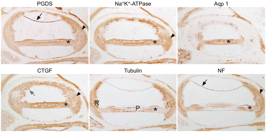
Fig. 2.
Celloidin removal with clove oil followed by immunostaining. All six panels show similar nonspecific staining pattern, regardless of antibody used. Reissner’s membrane (black arrows) shows background staining with PGDS and NF. Supporting cells (asterisks) show similar staining patterns with all six antibodies. Staining in spiral ligament (arrowheads) is diffusely brown with most of antibodies. In CTGF-stained tissue, bits of celloidin (gray arrow) are still evident in section near stria vascularis. Tubulin antibody starts to stain expected structures such as root cells (R) and pillar cells (P), but there is also great deal of brown background staining in section. Calibration bar — 50 µm.

