
Fig. 3.
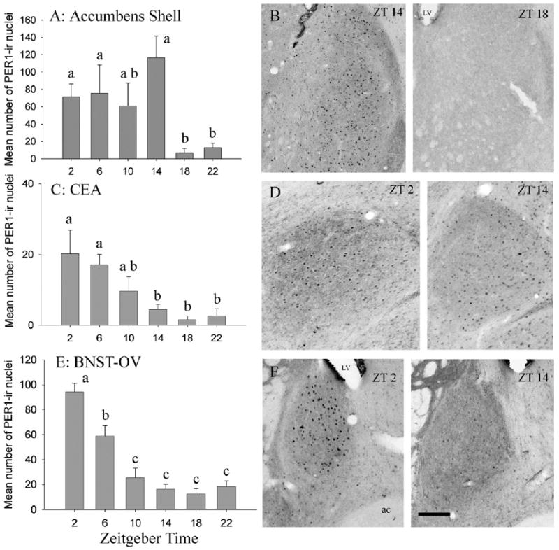
PER1 rhythm in the NA, the CEA, and the BNST-OV. Mean (±S.E.M.) numbers of cells containing PER1 and representative photomicrographs for the shell of the NA (A, B), the CEA (C, D), and the BNST-OV (E, F). LV, lateral ventricle; ac, anterior commissure. Scale bar=100 μm. Other symbols as in Fig. 2.