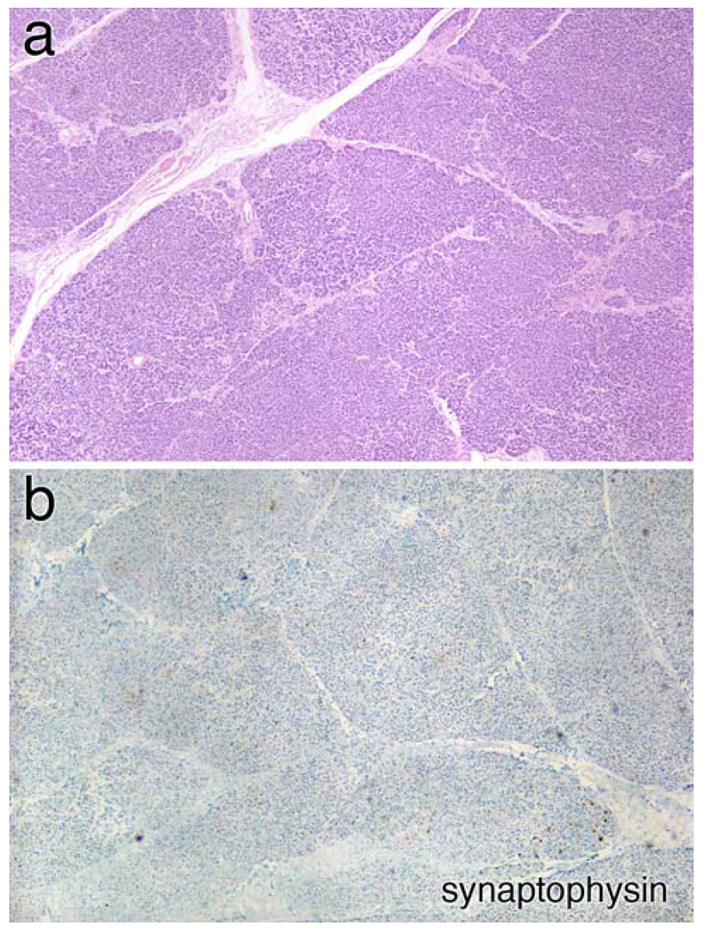
Figure 1.
Pancreas pathology. a Medium magnification view of the pancreas stained with hematoxylin and eosin (H&E). The organ has a normal lobular architecture; however no small, pale islands containing islets cells are visible. This is confirmed in panel (b), which has been immunostained with synaptophysin; no islands of islets cells are identified

