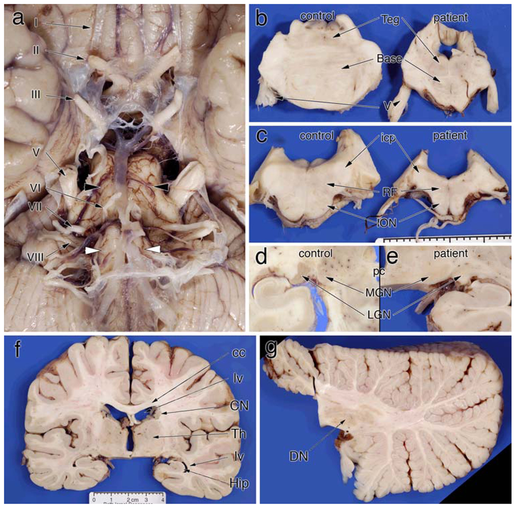
Figure 2.
Gross pathology. a Ventral view of the brainstem and optic nerves. The optic nerve (II) has a light brown color, rather than white, and has shrunken almost to the size of the oculomotor nerve (III). The remaining cranial nerves are unremarkable (IV–VIII). However, both the medulla (white arrowheads) and pons (black arrowheads) are significantly shrunken. b The diminution of the pontine base compared to a normal control. While the tegmentum (teg) has a normal height, the base is significantly smaller in all dimensions. c Similarly illustrates changes in the rostral medulla at the level of the inferior cerebellar peduncle (icp). The inferior olivary (ION) bulge is flattened and the medullar reticular formation (RF) is small. d A normal lateral geniculate nucleus (LGN) and medial geniculate nucleus (MGN) at the level of the posterior commissure (pc). e In the patient, the MGN has the same light gray color as the control; in contrast, the LGN completely lacks the darker brown color of the control. The coronal section in (f), which includes the LGN in (e), illustrates the essentially normal telencephalon. The lateral ventricles (lv) are not dilated, the corpus callosum (cc) is not atrophic or discolored, the thalamus (Th) is of normal size and configuration (except for the pallor in the LGN), and no hippocampal (Hip) shrinkage is present. Similarly, the radial section of the cerebellum in (g) displays no atrophy of the folia, and the dentate nucleus (DN) has a normal size and color

