Figure 4.
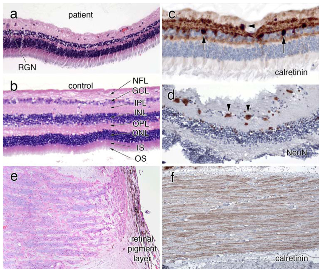
Eye. a, b Compare the retinal histology (H&E) in the patient and a control retina. They are at the same magnification, as determined by the similar width of the rod inner and outer segment layers. The patient’s retina has very few remaining retinal ganglion neurons (RGN) in the ganglion cell layer (GCL). Neurons in the inner (INL) and outer (ONL) neuronal layers are relatively preserved, while both the inner (IPL) and outer (OPL) plexiform layers are greatly diminished. c Stained for calretinin and shows an occasional RGN in the GCL (black arrowhead). Some calretinin-positive cells lie within the inner aspect of the INL. The Neu-N immunostain in (d), which reacts with a subset of neurons, reveals more RGN (black arrowheads), several of which are small or shrunken. e is an H&E-Luxol fast blue (H&E/LFB) stain of the head of the optic nerve. Many of the ganglion neuron axons have degenerated, resulting in pale myelin staining. Calretinin immunostains in (f) highlight remaining retinogeniculate axons

