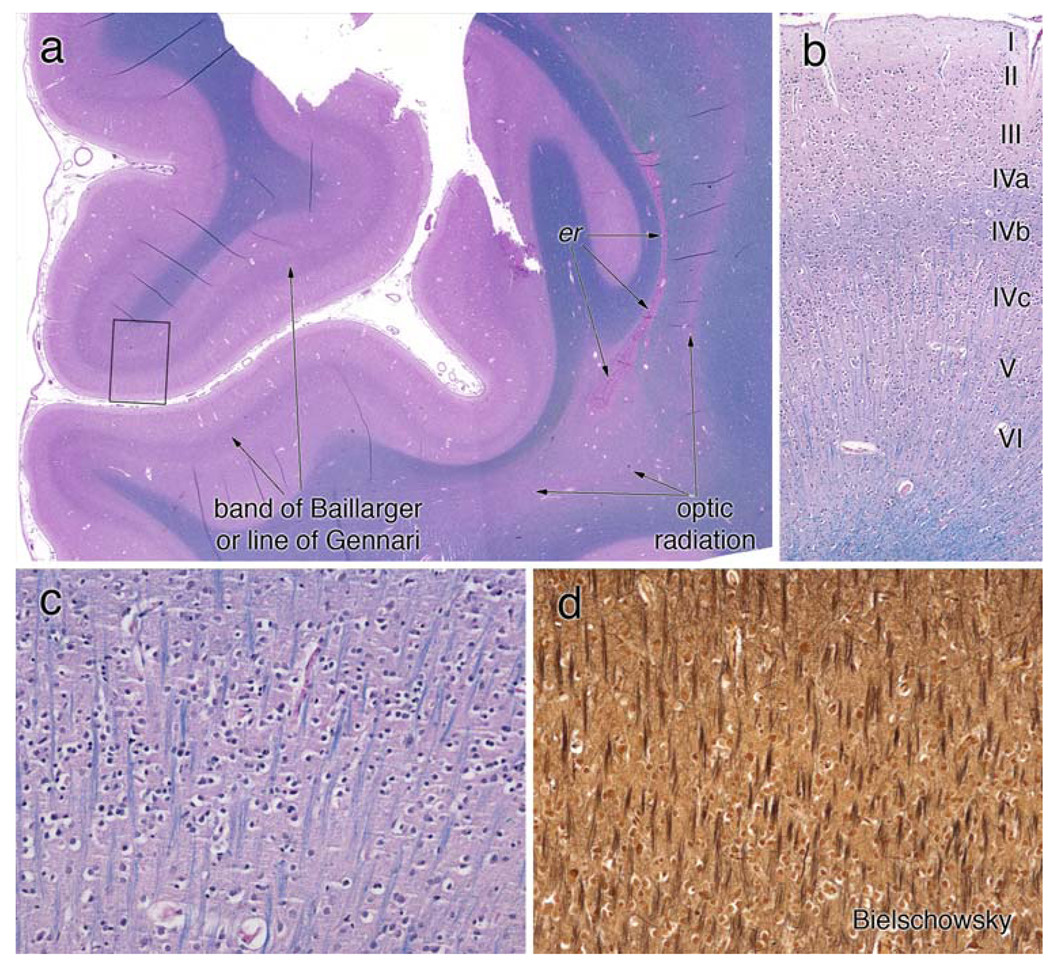
Figure 6.
Primary visual cortex. The coronal section of the primary visual cortex, stained with H&E/LFB, is illustrated in a field view in (a). The prominent white matter band within area 17 on gross inspection is composed of well-myelinated axons (line of Gennari or outer band of Baillarger). The optic radiation, which arose from the LGN and traveled lateral to the occipital horn of the lateral ventricle, exhibits pale myelin staining in this section, in keeping with the loss in the LGN. In this patient, the posterior recesses of the lateral ventricle had closed and left a line of ependymal rests (er). The small box in (a) is magnified in panel b to show the cortical layers. The outer band of Baillarger, representing intracortical connections, is well myelinated in layer IV-B. Small stellate or granular neurons in IV-C are present. Layer IV-C is illustrated at high magnification in (c) and (d). Radially oriented myelinated axons are identified on both the LFB (c) and Bielschowsky (d) stains. Neither stain shows myelin ovoids or axonal spheroids. Layer IV-C (and less so layer IV-A) is the major input lamina for the LGN; in this patient within the scope of this analysis, they are histologically normal

