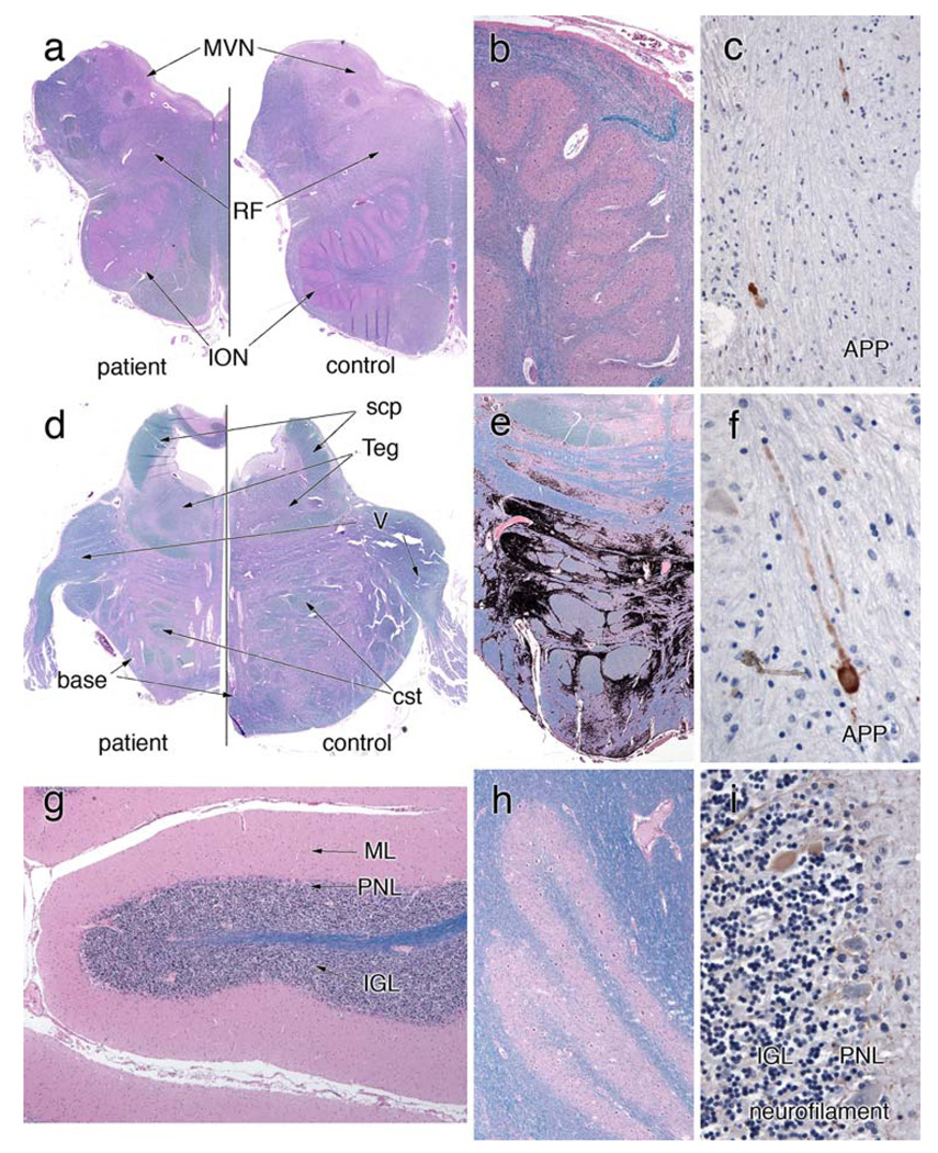
Figure 8.
Olivopontocerebellar system. a An H&E/LFB-stained section that compares the patient’s rostral medulla with a control. The patient’s entire medulla is small. Volume has been lost in the inferior olivary nucleus (ION) and reticular formation, and the medial vestibular nucleus (MVN) is slightly red, which represents a gliotic reaction to neuron loss. A higher magnification view of the ION in (b) reveals many remaining neurons; however, they are at a lower density than normal (see “Results”). They appear more spread out and do not show large patches of loss. Rare axons immunostain for amyloid precursor protein (APP) in (c). Similar to the medulla, the pons from the patient in (d) is small compared to the control. The tegmentum (Teg) has nearly the same size in both, but the base is moderately shrunken. The trigeminal nerve (V) is similar in both. To illustrate the patchy pattern of loss in the pontine base, the more gliotic regions were color-selected and then blackened (e). In these patches of gliosis, neurons are lost; in adjacent gray matter regions, they are spared. Notice that the dorsal aspect of the pontine base displays only slight involvement. As in the medulla, a few APP-immunoreactive axons are present in the pontine base white matter (f); these are rare. g, h H&E/LFB stains that illustrate the normal histology of the cerebellar gray matter. The molecular layer (ML) is not atrophic, the Purkinje neurons layer (PNL) has an appropriate number of large neurons, and the internal granular layer is normal. The dentate nucleus has a normal contingent of neurons. Correlating with the normal dentate nucleus, the superior cerebellar peduncle (scp) in (d) is fully myelinated, compared to the control. A neurofilament immunostain in (i) highlights only very rare axonal torpedoes in the IGL (brown-staining oval)

