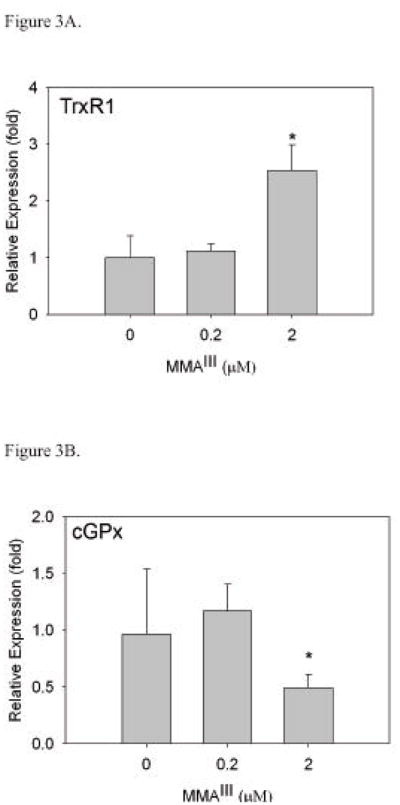
Figure 3. Exposure of WI-38 cells to MMAIII results in increases in TrxR1 mRNA levels.
Cells were cultured with MMAIII for 24 hours after which cells were harvested for real-time RT-PCR analysis. β-actin was used as an internal standard. Relative expression plotted is a representative experiment (cultures in triplicate) with standard deviation as error. Statistical significance was determined by t-test. There is a significant difference in TrxR1 mRNA levels (*, p<0.05) in WI-38 cells when treated with 2 μM MMAIII (A). As TrxR1 mRNA levels are increasing with exposure to MMAIII, cGPx levels significantly decrease with treatment of 2 μM MMAIII (*, p<0.05) (B).

