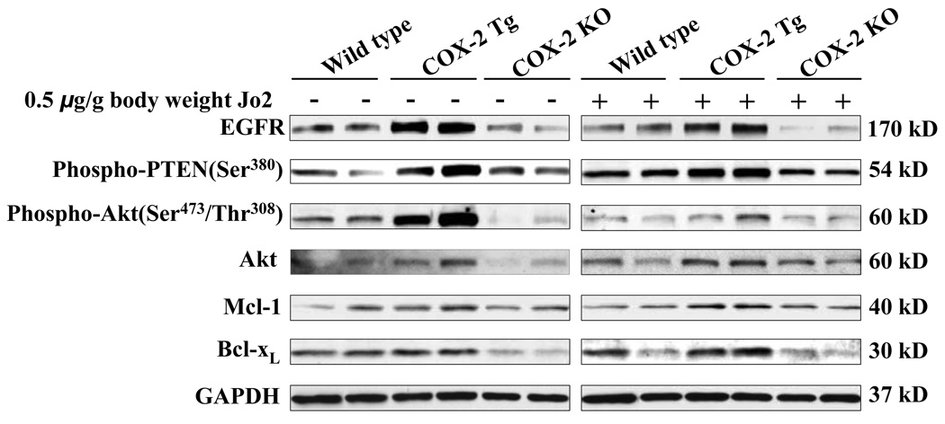
Figure 6. Changes of EGFR and related signaling molecules in mice with altered expression of COX-2.
The COX-2 Tg, COX-2 KO and wild type mice were injected intraperitoneally with saline or Jo2 (0.5 µg/g body weight). The livers were harvested 4 hours after the injection and the liver tissues were then homogenized. The obtained cellular proteins were subjected to SDS-PAGE and Western blot analysis to determine the protein levels of EGFR and its related signaling molecules, including phospho-PTEN, phospho-Akt, Akt, Mcl-1, and Bcl-xL. Western blot for GAPDH was shown as the loading control. The blots in this figure were obtained from two individual mice for each group.

