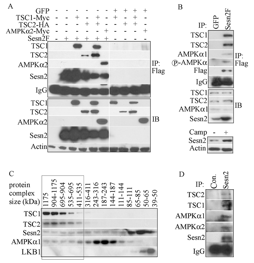
Figure 4. Sestrins interact with TSC1:TSC2 and AMPK.
(A) Co-immunoprecipitation of TSC1, TSC2 and AMPKα2 with Sesn2. HEK293 cells were co-transfected with Sesn2F or GFP expression vectors along with tagged TSC1, TSC2, TSC1 plus TSC2, or AMPKα2 plasmids as indicated. Sesn2F was immunoprecipitated with anti-Flag and presence of the indicated proteins in the immunecomplexes (IP) and total lysates (IB) was examined by immunoblotting. (B) Co-immunoprecipitation of endogenous TSC1, TSC2 and AMPKα1 with Sesn2. Sesn2F or GFP were induced in MCF7-tet OFF Sesn2F or GFP cells by incubation in low doxycycline concentration (0.01 µg/ml). After 24 hrs the cells were lysed. Sesn2F was immunoprecipitated with anti-Flag and presence of the indicated proteins in the immunoprecipitates (IP) and original lysates (IB) was examined. For comparison, MCF7 cells were incubated with camptothecin (Camp) for 12 hrs to induce Sesn2 expression that was examined by immunoblotting of the same amount of cell lysates as above. (C) Sesn2 co-elutes with TSC1, TSC2 and AMPKα in high molecular weight fractions. Extracts of H1299 cells were separated on a Superdex 200 gel filtration column and analyzed by immunoblotting. The elution positions (in kDa) of molecular weight standards are indicated at the top. (D) Co-immunoprecipitation of TSC1, TSC2 and AMPKα with endogenous Sesn2. Two months old wt male mice were injected with DEN (100 mg/kg). After 24 hrs, the liver was removed, homogenized and filtered. The lysate was separated by gel filtration as above and the high molecular weight fractions were combined and immunoprecipitated with anti-Sesn2 or control anti-rabbit (Con.) IgG. Presence of the indicated proteins in the immuneprecipitates was analyzed by immunoblotting.

