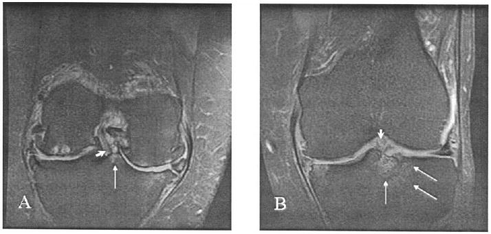
Figure 1.

Two types of central bone marrow lesion, as shown in coronal, fat-suppressed, T2-weighted magnetic resonance images. A, An anterior cruciate ligament (ACL) insertion (arrowhead) bordered by a grade 1 posterior tibial type 1 lesion (arrow). Bicompartmental tibiofemoral cartilage loss and osteophytes, subchondral bone cysts in the lateral femoral condyle, bone marrow edema in the medial tibia and femur, and partial maceration of both menisci are also shown. B, An ACL insertion (arrowhead) bordered by a grade 2 anterior tibial type 2 lesion with lateral extension (arrows). Mild cartilage thickening and small osteophytes in the medial tibial plateau and femoral condyle, bone marrow edema of the medial tibial plateau, and partial maceration and subluxation of the medial meniscus are also shown.
