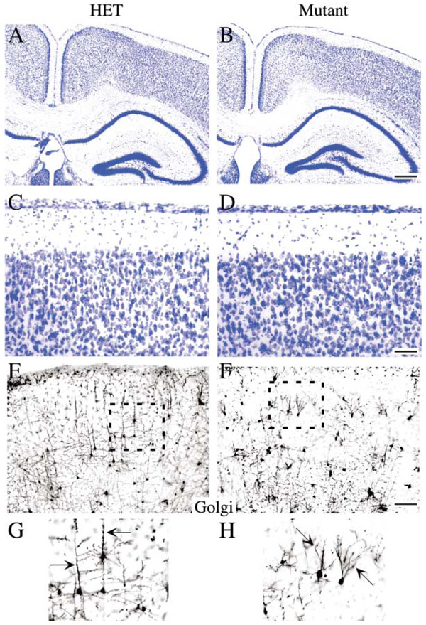
Figure 6. fak Deletion in Neurons Is Not Sufficient to Induce Cortical Dysplasia but Does Induce Altered Dendritic Morphology.
Coronal sections (40 µm) from adult control and nex-Cre/fak mutant forebrain stained with Nissl are indistinguishable at the gross anatomical level (A–D). The marginal zone was intact with no ectopic clusters of cells, and the hippocampus exhibited a normal morphology. However, Golgi staining of coronal sections (100 µm) did reveal abnormalities in dendritic branching in nex-Cre/fak mutants. Pyramidal neurons from layer III and V have a prominent apical dendrite extending perpendicular to the brain surface in control cells (arrow, [G]) that is notably perturbed in the fak−/− mutant (arrow, [H]). Scale bar, 500 µm (B) and 50 µm (D and F).

