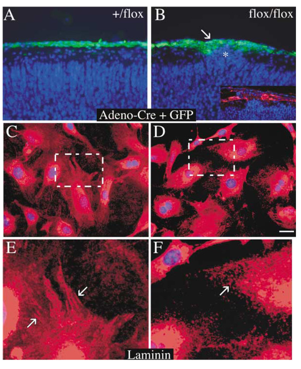
Figure 7. Adenoviral Recombination of Meningeal Fibroblasts Is Sufficient to Induce Cortical Dysplasia.
(A and B) Paraffin sections (7 µm) from E18.5 embryo heterozygous (+/flox, [A]) or homozygous (flox/flox, [B]) cortex following in utero injection of an adenovirus bearing Cre recombinase and GFP at E12.5/13 and stained with DAPI (blue) and anti-GFP (green). Cortical ectopias were present in the marginal zone of the flox/flox animal and coincided with interrupted laminin staining of the cortical basement membrane (inset, [B]).
(E–H) Laminin staining of primary meningeal fibroblasts isolated from E15 (flox/flox) and infected with either an adenovirus expressing GFP alone (E and G) or GFP plus Cre recombinase (F and H). fak−/− fibroblasts were unable to organize laminin into fibrillar structures as in control. Scale bar, 50 µm (B) and 25 µm (A–D).

