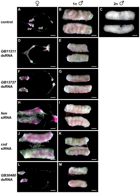
Figure 2. Reproductive organ development of 5th instar male and female larvae in the repression analysis of SDL genes.
(A–C) Reproductive organ development of untreated individuals: (A) A pair of normally developed ovaries (ov) and oviducts (od) from an untreated female. (B) A pair of normally differentiated testes from untreated haploid males consisting of densely packed layers of folded testioles. The paired spermducts are not shown. (C) A pair of normally differentiated testes from untreated diploid males consisting of less densely packed layers of folded testioles. The paired spermducts are not shown. (D–G) Repression analysis of gene GB11211 and GB13727. Normally developed gonads of females and haploid males injected with dsRNA devoted to repress the function of gene GB11211 (D–E) and GB13727 (F–G). (H–I) Repression analysis of the fem gene. (H) Pair of underdeveloped testes from a female treated with fem siRNA. The testes of this female individual are covered with oversized epithelial sheaths. The testioles are reduced in length and number when compared with the haploid (B) or diploid (C) males or the pseudomales after csd siRNA injection (J). The shape and course of spermducts appear normal. (I) Normally developed testes from a haploid male injected with fem siRNAs. (J–K) Repression analysis of the csd gene. (J) Pair of fully developed testes from a female treated with csd siRNAs. The number, length, and arrangement of testioles resemble entirely of those dissected from diploid males (C). (K) Normally developed testes from a haploid male injected with csd siRNA. (L–M) Repression analysis of the GB30480 gene. Normally developed gonads of females (L) and haploid males (M) injected with GB30480 dsRNA. Gonads were stained with aceto-orcein (reddish colouring of gonads), which facilitated the dissection process. Scale bars, 1 mm.

