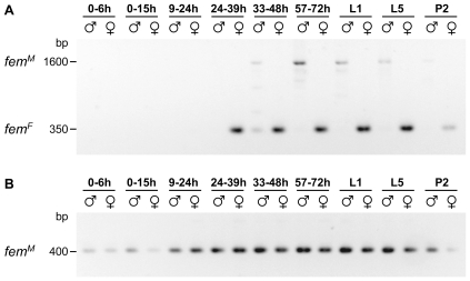
Figure 5. Developmental profile of fem mRNA expression.
Fragments corresponding to female (A) and male (B) fem mRNAs were independently amplified by RT-PCR and resolved by agarose gel electrophoresis. The weak ∼1,600 bp fragments observed in reactions devoted to amplify the female-specific fragment correspond to the male mRNAs. Differences in the amount of cDNAs in the different samples were adjusted prior to PCR amplifications. For the embryonic stages the hours after egg deposition are indicated. The early blastoderm is formed ∼12 h after egg deposition. L1 and L5 are 1st and 5th instar larvae, P2 are pupae at medium stage.

