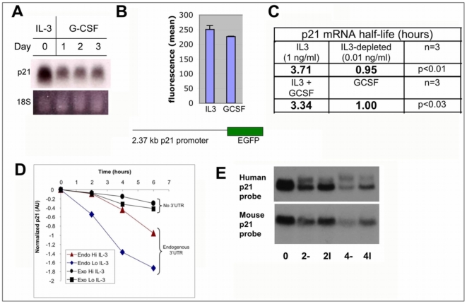
Figure 1. Posttranscriptional p21 mRNA regulation by IL-3.
A) Downregulation of p21 mRNA in differentiating myeloblasts. Northern blot analysis of total RNA from 32Dcl3 cells propagating in IL-3 (1 ng/mL) or following treatment with G-CSF (100 ng/mL) for indicated times. Cells remained viable over this time course. RNA harvested daily was probed with 32P-labeled murine p21 cDNA. Ethidium bromide stained 18S RNA is provided as a loading control. B) Comparable p21 promoter activity in proliferating and differentiating myeloblasts. 32Dcl3 cells stably transfected with EGFP driven by the murine p21 promoter were washed and either resuspended in IL-3 containing medium (1 ng/ml) or in G-CSF-containing medium (30 ng/ml) and EGFP expression measured 48 hours later by flow cytometry. Experiment was done twice with triplicate determinations. C) IL-3 stabilizes p21 mRNA. The half-life of p21 under indicated conditions was determined as described in Materials and Methods. p21 signals normalized against GAPDH on autoradiograms were plotted and mean decay rate of 3 independent experiments were determined. GAPDH remains stable under these conditions (Supplemental Table S1). D) IL-3-regulated turnover requires 3-UTR. Cells were stably transfected with p21 lacking UTR sequences, and decay of endogenous p21 (endo) compared to exogenous p21 (exo) cDNA was measured in chase experiments as above. E) Comparable decay of human and murine p21. Stable transfectants containing human p21 mRNA including the full-length 3′-UTR were exposed to medium with (+) or without (−) IL-3 for 2 or 4 hours after Actinomycin D as indicated, and mRNA blotted.

