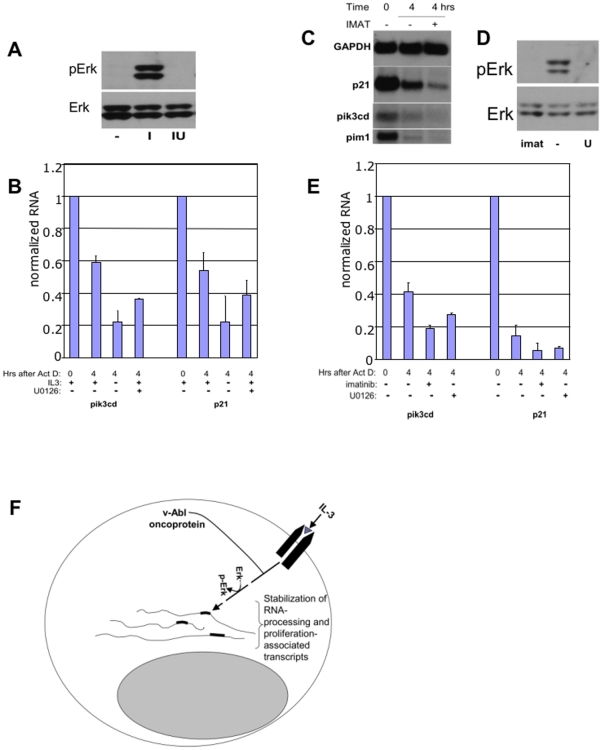
Figure 4. IL-3-mediated RNA stabilization involves MEK/ERK signaling.
A. IL-3 activates pErk in 32Dcl3 myeloblasts. Upregulation of phospho-Erk at 15 minutes after addition of IL-3 to 32Dcl3 myeloblasts washed free of IL-3 is evident (I). Pre-incubation for 20 minutes with the Mek1 inhibitor U0126 (at 10 uM) blocks this effect (IU), as does washout of IL3 (−). B. Downregulation of IL-3 induced transcript stabilization by Mek/ERK blockade. Cells were incubated with 10 uM of 10 uM of erk inhibitor U0126 concurrent with Actinomycin D. 1 ng/ml IL-3 (+) or vehicle was added 20 minutes later (time 0) and RNA was harvested 2 or 4 hours after IL-3 addition. p21 and pik3cd expression on Northern blots normalized to GAPDH is shown, with time 0 value set to 1.0 (n = 3). C. Imatinib inhibits mRNA stability in v-Abl 32Dcl3 cells. Imatinib (IMAT) or DMSO vehicle (−) were added following a 20 minute preincubation with actinomycin D, at time 0 as indicated. Cells were harvested for RNA at 0 and 4 hours and Northern Blots were probed with pik3cd, p21, pim1 and GAPDH. D. Erk phosphorylation. Phosphorylation of Erk in v-Abl transformants of 32Dcl3 is prevented by 15 minute incubation with imatinib (I, 5 uM) or with U0126 (U, 10 uM). E. Effects of imatinib and U0126 on transcript turnover. Data represents three separate experiments. Quantitation of RNA expression on Northern blots normalized to GAPDH, with time 0 value set to 1.0 is shown. F. Model of posttranscriptional pathway. Dashed line indicates possible involvement of additional signals.

