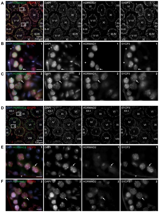
Figure 1. Testicular expression of HORMAD1 and -2 is restricted to meiotic germ cells.
Cryo-sections of adult testis were immunostained with anti-SYCP3 and either anti-HORMAD1 (A–C) or anti-HORMAD2 (D–F) antibodies. DNA was detected by DAPI. Overview of several immunostained tubules is shown in A and D. Epithelial cycle stages (Roman numerals) of testis tubules were determined based on SYCP3 localization pattern and DNA staining. HORMAD1/2 levels are highest in nuclei of early/mid pachytene cells (tubule stages I–IV) and decrease as cells progress to late-pachytene and diplotene (tubule stages VIII–XI). Bars are 100 µm in A and D, 10 µm in B, C, E and F. Boxes 1 and 2 from Panel A are shown at higher magnification in B and C. High HORMAD1 levels are detected in the nuclei of spermatocytes marked by chromosome axis component SYCP3. Axis-like staining of HORMAD1 is widespread across nuclei in zygotene (C,*) and diplotene (C, arrow). During pachytene, axis-like staining of HORMAD1 is restricted to a small chromatin domain, which appears to be the sex body based on DNA morphology (B, arrow). Boxes 1 and 2 from Panel D are shown at higher magnification in E and F. HORMAD2 is detected in the nuclei of spermatocytes marked by chromosome axis component SYCP3. Axis-like staining of HORMAD2 is widespread across nuclei in zygotene (F,*). During pachytene (E, arrow) and diplotene (F, arrow) the axis-like staining of HORMAD2 is restricted to a small chromatin domain, which appears to be the sex body. No specific accumulation of HORMAD1 and -2 is observed in sperm, spermatid and any testicular somatic cells, e.g.: Sertoli cells (arrowhead in B and E) and myoid cells (+ in B).

