
Figure 5.
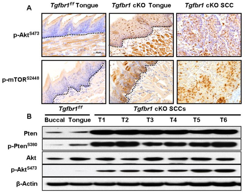
Activation of the PI3K/Akt pathway in Tgfbr1 cKO mice. A, Immunostaining revealed a significantly increased number of positive cells of p-Akt, p-mTOR in the SCCs that developed in Tgfbr1 cKO mice. The dotted lines delineate the adjacent epithelial compartment. Bar, 50 μm. B, A significantly increased level of unphosphorylated PTEN, an active form of the protein, was detected in all SCCs that developed in the DMBA-treated Tgfbr1 cKO mice. However, comparable elevated levels of the phosphorylated form of Akt (p-Akt) were also observed in SCCs by Western blot analysis.