
Fig. 4.
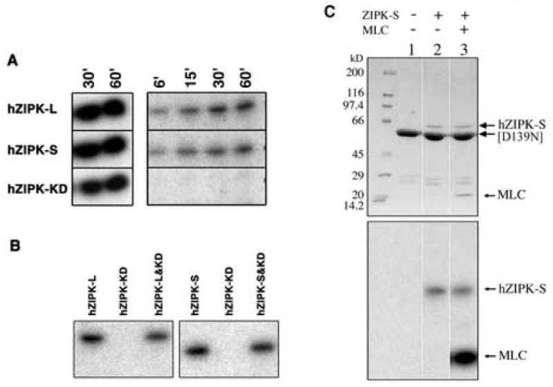
Autphosphorylation sites within the non-kinase domain of hZIPK-S. A. Autophosphorylation of hZIPK isoforms and hZIPL-KD. Autophosphorylation was done as described in MATERIALS AND METHODS. Left panel shows the autoradiogram of MLC20 phosphorylation by hZIPK. Right panel shows the autoradiogram of hZIPK autophosphorylation. B. Failure of phosphorylation of hZIPK-KD by hZIPK isoforms. Left panel: hZIPK-L (first lane), hZIPK-KD (second lane), or hZIPK-L + hZIPK-KD (third lane) were phosphorylated with 0.2 mM [γ32P]-ATP at 25°C for 15 min in the reaction buffer. Right panel: hZIPK-S (first lane), hZIPK-KD (second lane), or hZIPK-S + hZIPK-KD (third lane). Note that only hZIPK-L or-S but not hZIPK-KD was phosphorylated. C. Failure of phosphorylation of D139N hZIPK-KD by hZIPK-S. The hZIPK-KD (D139N) mutant was incubated with hZIPK-S in the presence of 0.2 mM [γ-32P] ATP at 25°C for 15 min. Upper panels show Coomassie Brilliant Blue (CBB) staining, and lower panels show autoradiography. Note that hZIPK-S phosphorylated MLC20 but not [D139N] hZIPK-KD.