
Figure 2. Superposition of MjaOgg on A) hOGG1 and B) Pa-AGOG.
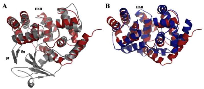
A) Structural superposition of unliganded hOGG1 (PDB ID code: 1KO9) (Ogg1; gray) on MjaOgg (Ogg2; red) illustrating the similar architecture of domains B and C of hOGG1 to the corresponding domains (N- and C-terminal) in Ogg2 and the absence of the hOGG1 A domain in Ogg2. B) Structural superposition of Pa-AGOG (PDB ID code: 1XQO) (AGOG; blue) onto MjaOgg (Ogg2; red) showing a similar fold for the two enzymes.