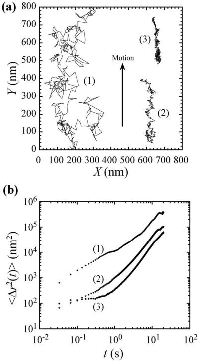
FIG. 1.
Particle tracking microrheology with 100 nm diameter fluorescent polymer spheres embedded within NIH3T3 fibroblast cells. (a) 20 s trajectories of typical transport in NIH3T3 cells. The approximate speeds of particles (2) and (3), calculated by end-to-end displacement, are 18 and 12 nm/s, respectively. Particle 1 shows a partial trajectory that continues past the top of the figure. (b) Time-dependent mean-square displacements (MSDs) of the particles in (a). All the particles exhibit directional motion correlated to cell crawling (arrow).

