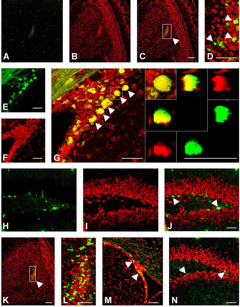
Fig. 10.
Colocalization of CXCR4 (A–J) or CCR2 (K–N) expression with Ki67 in neurogenic regions of 5-week-old mouse brain. FISH was performed using CXCR4 or CCR2 antisense probes in conjunction with immunostaining with a Ki67 antibody. Both CXCR4 and CCR2/ CCR5 colocalized with Ki67 in the olfactory ventricle (arrowheads in picture obtained at higher magnification, D: CXCR4, L: CCR2) of the olfactory bulb (OB), in the subventricular zone (SVZ) (arrowheads in G: CXCR4 and M: CCR2), and in the subgranular zone (arrowheads in J: CXCR4 and N: CCR2) of the dentate gyrus (DG). The right panels inserted in G shows the colocalization of CXCR4 and Ki67 at higher magnification in the SVZ. The panel at the left corner shows a xy-projection of a z-stack, Ki67 in green and CXCR4 in red. Colocalization of CXCR4 and Ki67 was confirmed by 3D reconstitution. Left and bottom panels illustrate the reconstructed view in xz plane and top and right panels show the yz plane. Both xz- and yz-reconstructions are also displayed in split channel mode to allow further assessment of the double labeling. Scale bars = 100 µm.

