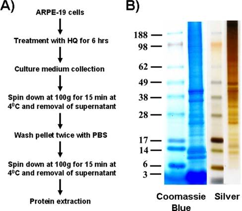
Fig. 2.
Isolation of blebs. A, scheme for bleb isolation. ARPE-19 cells were treated with HQ (100 μm) for 6 h. Culture medium was collected and centrifuged at 100 × g for 15 min at 4 °C. The resulting pellet was washed twice with PBS and resuspended. The resuspended pellet was centrifuged at 100 × g for 15 min at 4 °C, and the supernatant was removed. Blebs were collected and used for protein extraction. B, representative one-dimensional gel showing the Coomassie Blue and silver stainings of resolved proteins present in ARPE-19 blebs.

