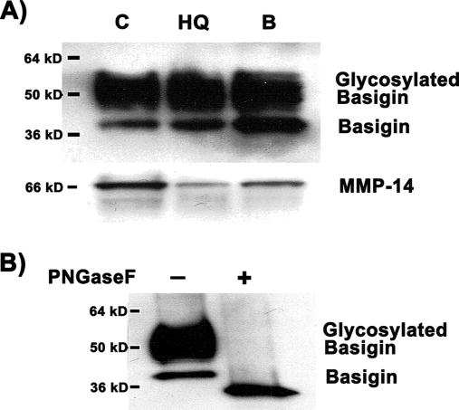
Fig. 4.
Western blot analysis of basigin and MMP-14 expression in hydroquinone-induced blebs. Protein expression was analyzed in 20 μg of total ARPE-19 cell lysate. A, basigin and MMP-14 expression in control, untreated cells (C), cells treated with 100 μm HQ for 6 h (HQ), and isolated HQ-induced membrane blebs (B). Basigin analysis shows a higher molecular mass, broad band corresponding to highly glycosylated species of basigin. B, Western blot analysis of basigin in isolated HQ-induced membrane blebs before and after digestion with the enzyme PNGaseF, which selectively removes N-linked carbohydrate residues. The blot from a representative experiment is shown. The number on the left represents the molecular mass of the protein.

