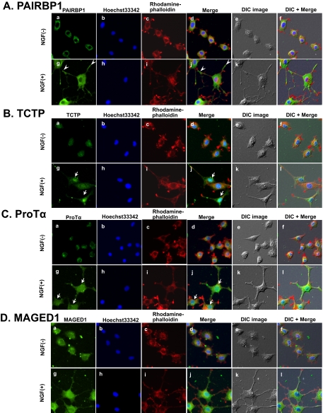
Fig. 5.
Analysis of NGF-responsive proteins by ICC. PC12 cells were treated with NGF for 48 h. Cells were fixed and incubated with antibodies against the indicated proteins (A, PAIRBP1; B, TCTP; C, ProTα, and D, MAGED1) followed by detection with Alexa Fluor 488-labeled secondary antibodies (a and g) and observation with a fluorescence microscope. Nuclear and F-actin were stained with Hoechst33342 (b and h) and rhodamine-phalloidin (c and i), respectively. The merged images for a, b, c and g, h, i are shown in d and j, respectively. The merged images for d, e and j, k are shown in f and l. Differential interference contrast (DIC) images of PC12 cells in the same field are shown in e and k. Arrowheads indicate the PAIRBP1 expression in the neurite junctions and tips. Arrows indicate the TCTP and ProTα expression in the nucleus.

