Fig. 6.
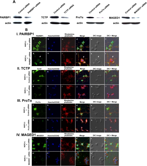
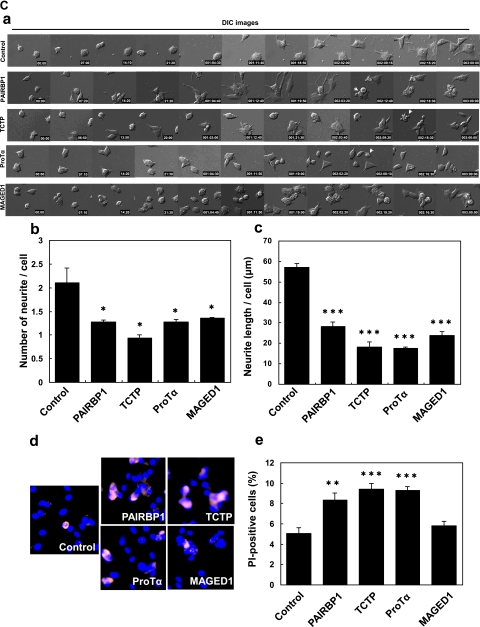
The effects caused by the suppression of the NGF-inducible proteins on PC12 cell differentiation. A, the cells were transfected with siRNA for each NGF-inducible protein or control siRNA for 24 h and then treated with NGF for 48 h. Immunoblot images were taken after treatment with each siRNA of the protein. Down-regulation of these proteins was confirmed. B, the cells were transfected with siRNA for each NGF-inducible protein or control siRNA for 24 h and then treated with NGF for 48 h. Cells were fixed and incubated with the antibodies against the indicated proteins (I, PAIRBP1; II, TCTP; III, ProTα; and IV, MAGED1) followed by detection with Alexa Fluor 488-labeled secondary antibodies and observation with a fluorescence microscope (a and g). Nuclear and F-actin were stained with Hoechst33342 (b and h) and rhodamine-phalloidin (c and i), respectively. The merged images for a, b, c and g, h, i, are shown in d and j, respectively. The merged images for d, e and j, k are shown in f and l, respectively. Differential interference contrast (DIC) images of PC12 cells in the same field were shown in e and k. The suppression of expression of each protein led to significant morphological changes, such as inhibition of neurite formation and induction of cellular aggregation in differentiating PC12 cells. Arrowheads indicate the PAIRBP1-suppressed cells. C, effects on neurite outgrowth and survival of PC12 cells of the siRNA treatment of the NGF-inducible proteins. a, representative time lapse images of NGF-stimulated PC12 cells treated with each siRNA for NGF-inducible proteins. The cells were transfected with siRNA for each NGF-inducible protein or control siRNA and stimulated with NGF for 72 h. A significant inhibition of neurite formation was observed due to each siRNA treatment. Arrows show the apoptotic phenotypes of PC12 cells. b and c, the cells were transfected with siRNA for each NGF-inducible protein or control siRNA and stimulated with NGF for 48 h under the condition of low serum (1% horse serum). The average of the number and total length of neurites of the PC12 cells are shown on the y axis (b and c). The data are expressed as means and S.D. of the three independent experiments (n = 3). For each experiment, more than 50 cells were counted. d and e, effects on PC12 cell survival of the siRNA treatment of the NGF-inducible proteins. The cells were transfected with siRNA for each NGF-inducible protein or control siRNA, stimulated with NGF for 48 h, fixed, and stained with PI and Hoechst33342 dye. Representative images of PI and Hoechst staining of PC12 cells treated with each siRNA and control and stimulated with NGF are shown (d). The percentage of PI-positive PC12 cells in total Hoechst-stained PC12 cells is shown on the y axis (e). The data and error bars are expressed as means and S.D. of the three independent experiments (n = 3), respectively. For each experiment, more than 500 cells were counted. *, p < 0.05; **, p < 0.01; and ***, p < 0.001 versus NGF + control siRNA treatment (Student's t test).


