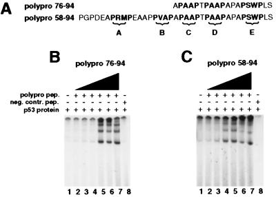
Figure 4.
Activation of p53 by synthetic polyproline peptides spanning residues 80–93. Sequences of the two peptides in single letter code. PXXP motifs, termed “A–E,” are depicted in boldface letters (A). EMSAs of full-length p53 with radiolabeled Ep21 oligonucleotide (B and C). Lanes 1 represent basal uninduced p53 DNA binding activity. In lanes 2–7 increasing concentrations of the indicated polyproline peptide had been added to the DNA binding reactions:0.1 mM (lanes 2); 0.25 mM (lanes 3); 0.5 mM (lanes 4); 1 mM (lanes 5); 2.5 mM (lanes 6); 7.5 mM (lanes 7). Lane 8: 0.75 mM of control peptide had been added to the reaction mixture.

