Fig. 4.
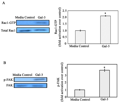
Gal-3 activates Rac1 GTPase and FAK in corneal epithelial cells. (A) HCLE cells were growth-factor-starved and were incubated in the presence or absence of Gal-3 in serum-free medium. Cell extracts were subjected to pulldown assays using GST-PAK to examine GTP-bound Rac1 (active Rac1) levels. Precipitated GTP-Rac1 as well as total cell lysates were examined by immunoblot analysis using a mouse anti-Rac1 mAb. The intensity of each band was quantified by densitometry and the ratios of GTP-loaded Rac1 to total Rac1 were determined and normalized to cells exposed to serum-free medium alone (media control). Note that the levels of total Rac1 (GTP-bound + GDP-bound forms) were similar regardless of whether the cells were exposed to Gal-3 or not. By contrast, within 30 seconds of exposure to Gal-3, there was a 2.3-fold increase in the amount of activated Rac1 (bar graph), and enhanced Rac1 activity was maintained up to 10 minutes (data not shown). Values are mean ± s.e.m. of three separate experiments. *P<0.01 compared to media control. (B) Electrophoresis blots were probed with anti-FAK or anti-Y397 phosphorylated FAK monoclonal antibodies. Note that FAK is activated (p-FAK lanes) after exposure to Gal-3, whereas total FAK levels (FAK lanes) are similar in the control and Gal-3-exposed cells. The intensity of each band was quantified and ratios of p-FAK to total FAK was calculated and normalized to control values (bar graph). *P<0.05 when compared with control; data are mean ± s.e.m. from three independent experiments.

