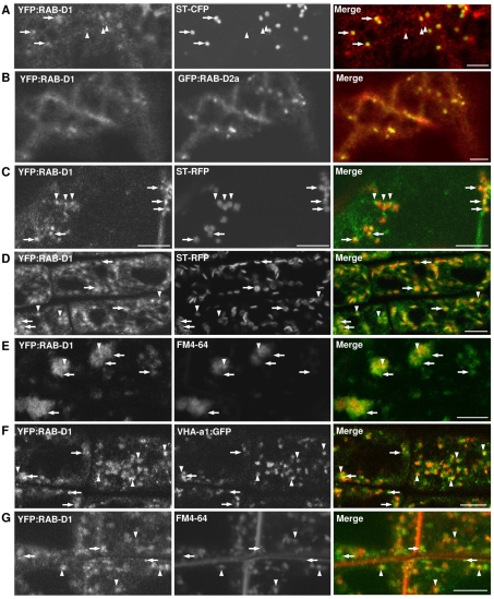
Fig. 1.
Localisation of YFP:RAB-D1. Confocal laser scanning microscope (CLSM) images of tobacco (A,B) or Arabidopsis (C-G) cells coexpressing YFP:RAB-D1 (green) with various markers (red). (A,B) YFP:RAB-D1 in tobacco leaf abaxial epidermis coexpressing either the Golgi marker ST-CFP (A) or GFP:RAB-D2a (B). (C) Arabidopsis leaf abaxial epidermis coexpressing the Golgi marker ST-RFP. (D-G) Localisation of YFP:RAB-D1 in Arabidopsis root tips together with the Golgi marker ST-RFP (D), the TGN marker VHA-a1:GFP (F), or the endosomal dye FM4-64 (E,G). Root tips in E were treated with brefeldin A. Arrows indicate YFP:RAB-D1-labelled punctate structures that associate with Golgi stacks. Arrowheads show structures that associate with the TGN. Scale bars: 5 μm.

