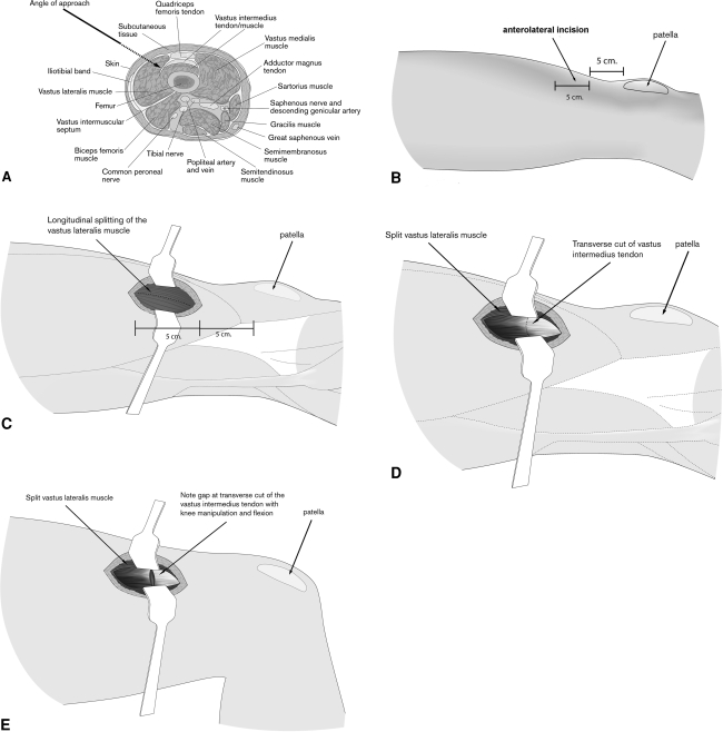
Fig. 1A–E.
The schematic diagrams show the surgical steps for limited quadricepsplasty. The (A) anatomy of the distal thigh, (B) location of the incision, (C) splitting of the vastus lateralis muscle, (D) exposure and incision of the vastus intermedius tendon/fascia, and (E) the separated vastus intermedius tendon/fascia are shown.

