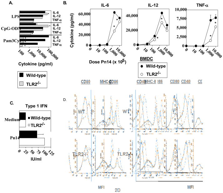
Fig 2. Analysis of in vitro cytokine secretion and phenotypic maturation of Pn14-stimulated TLR2−/− and wild-type bone marrow dendritic cells (BMDC).
BMDC from WT and TLR2−/− (both B6.129 background) mice were incubated with (A) LPS (2μg/mL), CpG (2μg/mL), Pam3CSK4 (150 ng/mL) or (B) varying doses of Pn14 for 24 h and the levels of IL-6, IL-12 and TNF-α were measured. The results are representative of two independent experiments. (C) BMDC from WT and TLR2−/− (both C57BL/6 background) mice were incubated with Pn14 for 24h and the levels of type I IFN were measured. The results are representative of two independent experiments. *represents significance (p<0.05) for A, B, and C, by Student’s t-test. (D) BMDC from WT and TLR2−/− (both B6.129 background) mice were stimulated with Pn14 for 24 h. Cell surface expression levels of MHC-II, CD80, CD86, and CD40 were measured by flow cytometry. Solid lines represent BMDC cultured in medium alone; dotted lines represent BMDC cultured with Pn14. One of two independent experiments is shown.

