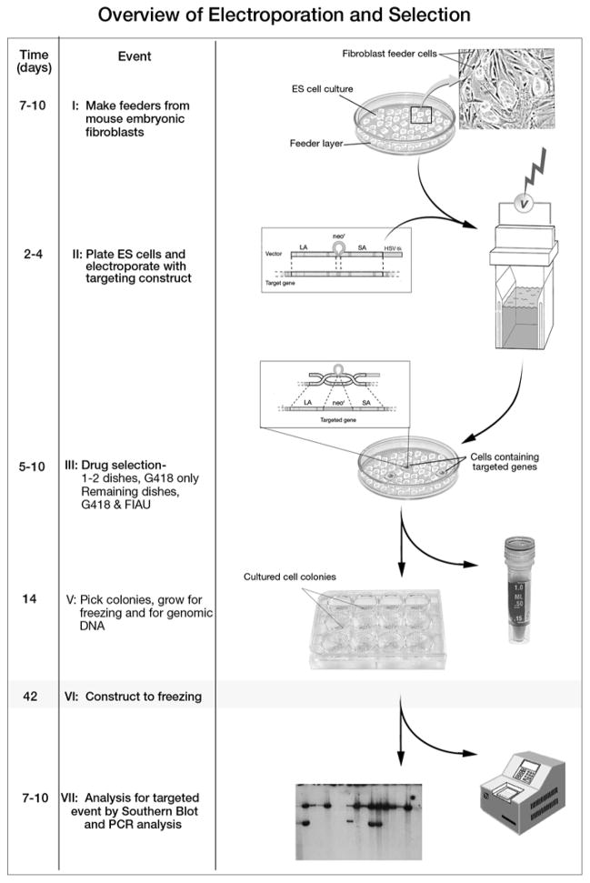
Figure 1.
Overview of Electroporation and Selection. The left column shows the approximate number of days at each critical step in the preparation of targeted embryonic stem cells. The middle column describes key procedural events. The last column is an illustration of each critical event. Note that in stages II through V embryonic stem cells should remain as close to pluripotent as possible. Once the cultured ES cells have been plated to the tissue culture plate depicted in step V, they may become differentiated. These cells will used for DNA screening as depicted in step VII. In step VII, the left panel represents an autoradiographic film image of a Southern blot. Southern blot is the definitive test for detection of a targeted event. Many investigators first screen all clone DNA samples by PCR followed by confirmation with Southern blot. A strategy for differentiating between the knockout allele and the wild-type allele should be worked out in advance of initiating a targeting experiment.

