
Fig. 4.
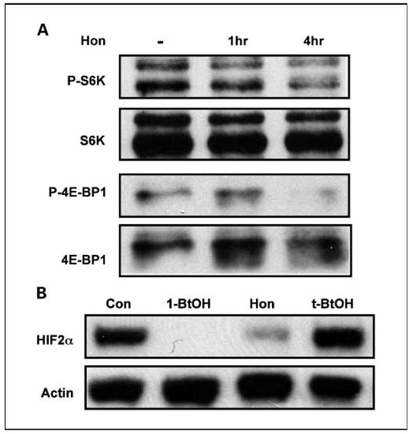
Honokiol suppresses downstream targets of PLD survival signals. A, MDA-MB-231 cells were plated in DMEM with 10% serum. Forty-eight hours later, the cells were shifted to 0.5% for 16 h. Honokiol (20 μmol/L) or control ethanol was then added for the indicated times. The cells were then harvested and analyzed for levels of S6K, phosphorylated S6K (P-S6K), 4E-BP1, and P-4E-BP1 by Western blot analysis as described previously (10). B, 786-O cells were plated and then shifted to medium containing 0.5% serum 24 h later. Eighteen hours later, the cells were treated with 1-BtOH (0.8%), honokiol (20 μmol/L), and t-BtOH (0.8%) as indicated. Cell lysates were prepared 4 h later and examined for HIF2α and actin expression by Western lot analysis. The experiment shown is representative of two independent experiments.