
Figure 5. TLR2 activation of CaMKII leads to phosphorylation of TBLR1.
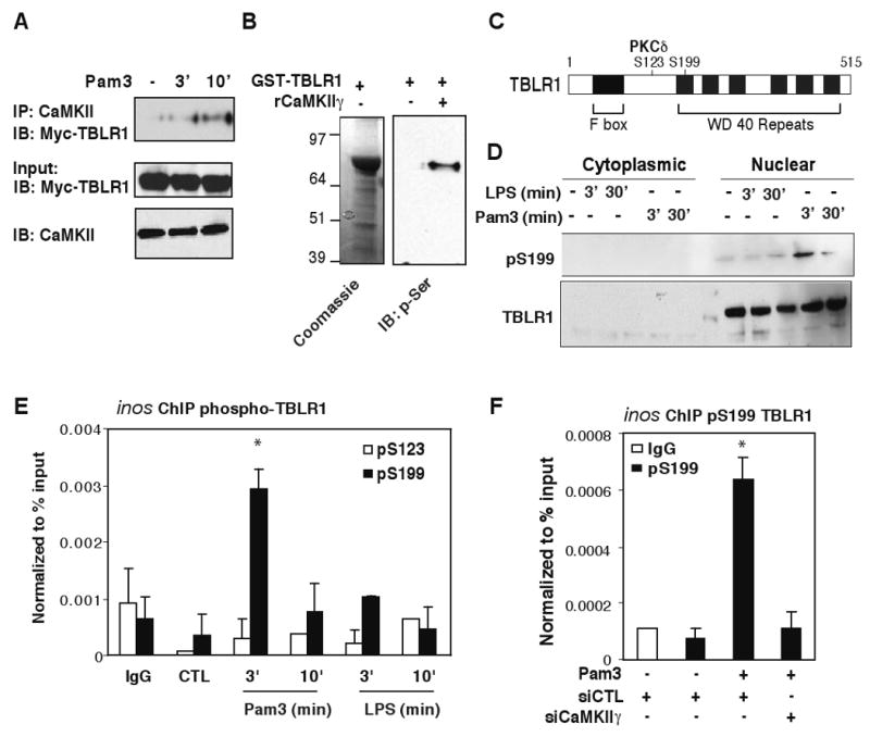
(A) RAW264.7 cells were transfected with Myc-TBLR1 expression construct and treated with Pam3 for 3 and 10 min. IP was carried out with CaMKII-specific antibodies and TBLR1 was detected by anti-Myc antibodies by IB. Bottom panel illustrates inputs for Myc-TBLR1 and CaMKII in each sample. (B) Recombinant GST-TBLR1 proteins were used as substrates for CaMKII in vitro. Bacterial GST-TBLR1 was captured on glutathione agarose and incubated with or without 0.5 μg of activated rCaMKIIγ. Anti-phospho-serine antibody was used to detect phosphorylated TBLR1 proteins by IB. (C) Diagram of TBLR-specific phosphorylation sites. (D) BMDM were challenged with LPS or Pam3 for 3 and 30 min. Cytoplasmic and nuclear extracts were immunoblotted for total and pS199 TBLR1. (E) ChIP assays for phospho-TBLR1 in BMDM treated with Pam3 or LPS for the indicated times using antibodies against p-S199-TBLR1 or p-S123-TBLR1. (F) ChIP assays for p-S199-TBLR1 on the inos promoter in BMDM transfected with siCTL or siCaMKIIγ siRNAs and challenged with Pam3 for 3 minutes. *p<0.05 versus non-treated controls. Errors bars represent SD.