
Fig. 4.
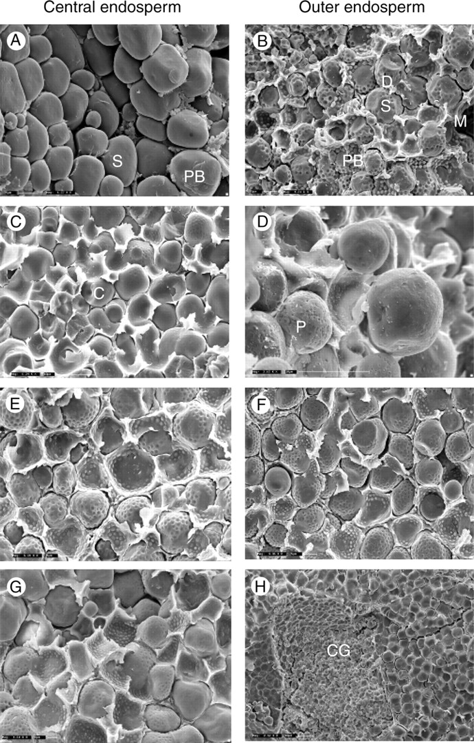
The left-hand column shows the variation in the central endosperm and the right-hand column compares the outer layers. (A, B) Representative images of S. bicolor, showing the standard floury and vitreous endosperm, respectively. (C–H) Images from outside the Eu-Sorghums are representative of the variations observed across the species. PB, Protein bodies; M, matrix; S, starch granule; D, indentations left by protein bodies; C, channels; P, pores; CG, small polygonal starch granules forming compound granules.