Fig. 3.

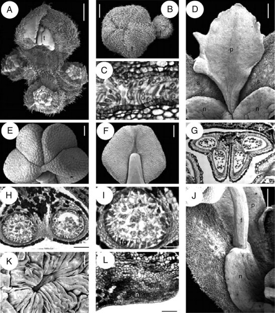
Quillaja saponaria, flower and inflorescence structure. A, B, D–F, J and K are SEM micrographs; C, G, H, I and L are transverse sections. (A) Young flowering branch with terminal bud and surrounding lateral buds. (B) Floral buds with hairy valvate calyx. (C) Sepals postgenitally united by cohesive hairs. (D) Petal at anthesis attached to a nectary sinus and alternating with sepals. (E, F) Top and dorsal view of anther. (G) Stamen transverse section with epidermis and connective with tanniniferous cells. (H) Theca and connective tissue. (I) Transverse section of sporangium with secretory parietal tapetum and tetrahedral tetrads. (J) Nectary lobe united with the opposite sepal and attached to a stamen. (K) A single stomata from a nectary. (L) Transverse section of sepal (above) and nectary (below). f, filament; l, lateral flower; n, nectary; p, petal; s, sepal; t, terminal flower; tp, tapetum. Scale bars: A, B, D, J = 0·5 mm; C, K = 20 µm; E, F = 250 µm; G = 200 µm; H, L = 100 µm, I = 50 µm.
