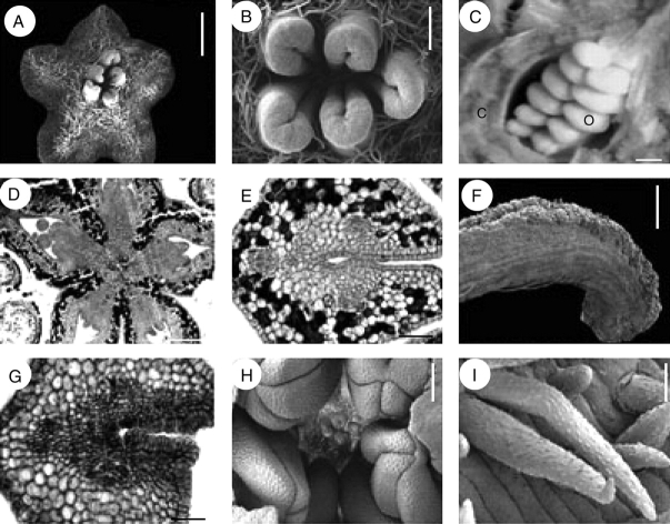
Fig. 4.
Quillaja saponaria, floral structure. A, B, F, H and I are SEM micrographs; C picture under stereoscope; D, E and G transverse sections. (A) Top view of gynoecium. (B) Distal and glabrous part of gynoecium. (C) Rows of ovules attached at the marginal side of a carpel; carpel wall partially removed. (D) Symplicate area of gynoecium in transverse section surrounded by layers of tanniniferous tissue. (E) Transverse section, style below stigmatic area displaying three main vascular bundles, and PTT covering ventral furrow. (F) Exposed stigmatic zone at anthesis. (G) Transverse section of style at stigmatic area level with secretory cells covering ventral furrow. (H) Fully developed anthers surrounding underdeveloped gynoecium. (I) Indumentum covering inflorescence axes, sepals and gynoecium, with warty ornamentation. c, carpel; o, ovule. Scale bars: A, C, H = 0·5 mm; B = 150 µm; D = 200 µm; E, G = 50 µm; F = 250 µm; I = 10 µm.

