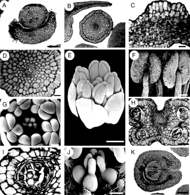
Fig. 7.

Suriana maritima, floral structure. E–G and J are SEM micrographs; A–D, H, I and K are transverse sections. (A) Petal surrounded by staminode base. (B) Petal and filament. (C) Petal, dorsal side at the upper side of the figure. (D) Filament. (E) Oblique view of bud at mid-development. (F) Dorsal view of basifixed, mature, X-shaped anthers. (G) Polar view of floral bud showing gynoecium and androecium during mid-development. (H) Transverse section of an anther. (I) Sporangium with parietal tapetum. (J) Lateral view of apocarpous, pentamerous gynoecium during mid-development. (K) Proximal side of carpel showing marginal placentation of ovules. c, carpel; f, filament, p, petal; stm, staminode. Scale bars: A, B, H, K = 100 µm; C = 10 µm; D, I = 20 µm; E = 0·5 mm; F, G, J = 200 µm.
