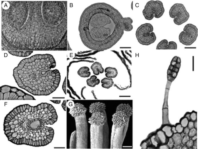
Fig. 8.
Suriana maritima, floral structure. G is an SEM micrograph; A–F and H are sections. (A) Transverse section of a carpel displaying the integument and obturator projections. (B) Transverse section of the distal part of ovules; note the carpel filled with mucilage. (C) Transverse section of styles at the level below the anthers; stylar canal is isolated from the exterior at the ventral side. (D) Style at the level of the anthers, with stylar canal filled with mucilage. (E, F) Styles above anther level displaying closed stylar canal surrounded by PTT and filled with mucilage. (G) Side view of group of mature papillate stigmas. (H) Glandular hair on sepal. c, carpel; i, integument; o, obturator; ov, ovule; sy, style. Scale bars: A, D = 50 µm; B, C = 100 µm; E, G = 200 µm; F, H = 20 µm.

