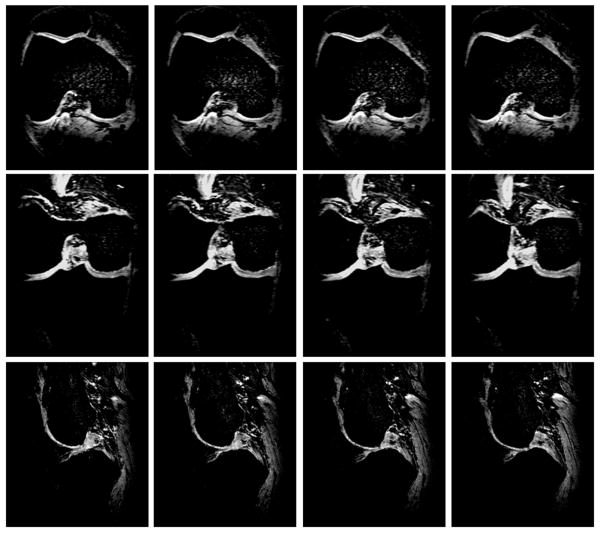
Fig. 9.
Four representative consecutive proton T1-weighted images of human cartilage from an OA subject in three orthogonal directions of axial (top), coronal (middle), and sagittal (bottom) at 7T, respectively. Acquisition parameters: 3D-FLASH, fat suppression, TR/TE = 20 ms/4.3 ms, Flip Angle = 100, Averages = 1, Bandwidth = 130 Hz/pixel, thickness = 1 mm, FOV = 100 mm × 100 mm, Matrix = 512 × 512.

