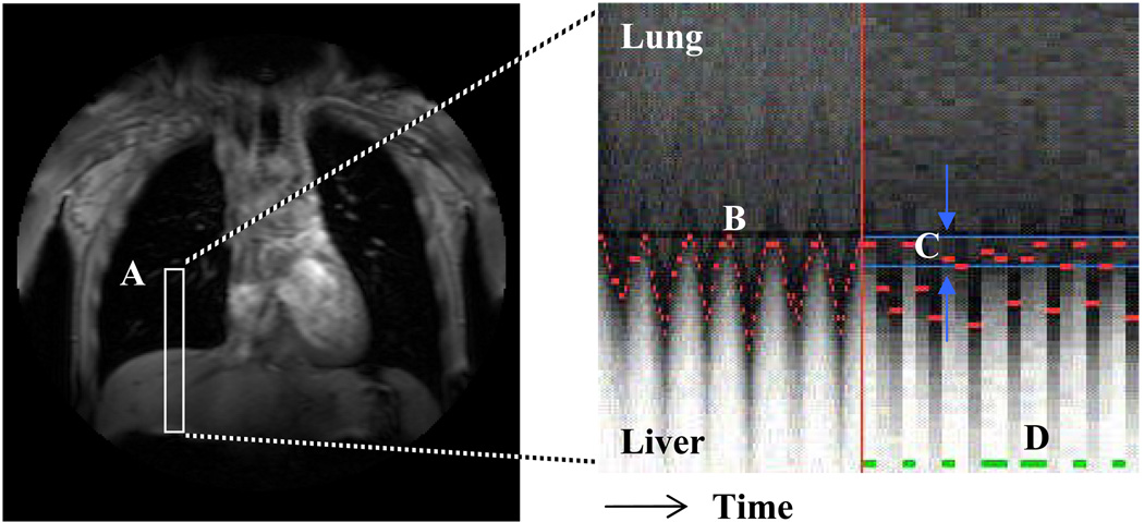
Figure 1.
A one-dimensional navigator-echo pulse (A, in white) is positioned over the liver and right lung to track the motion of the diaphragm over time. The boundary between the hypo-intense lung and hyper-intense liver at each heartbeat is recognized and tracked over time (B, in red). An acceptance window is defined at end respiration (C, in blue) and scan data is acquired for heartbeats during which the diaphragm lies within the acceptance window (D, in green).

