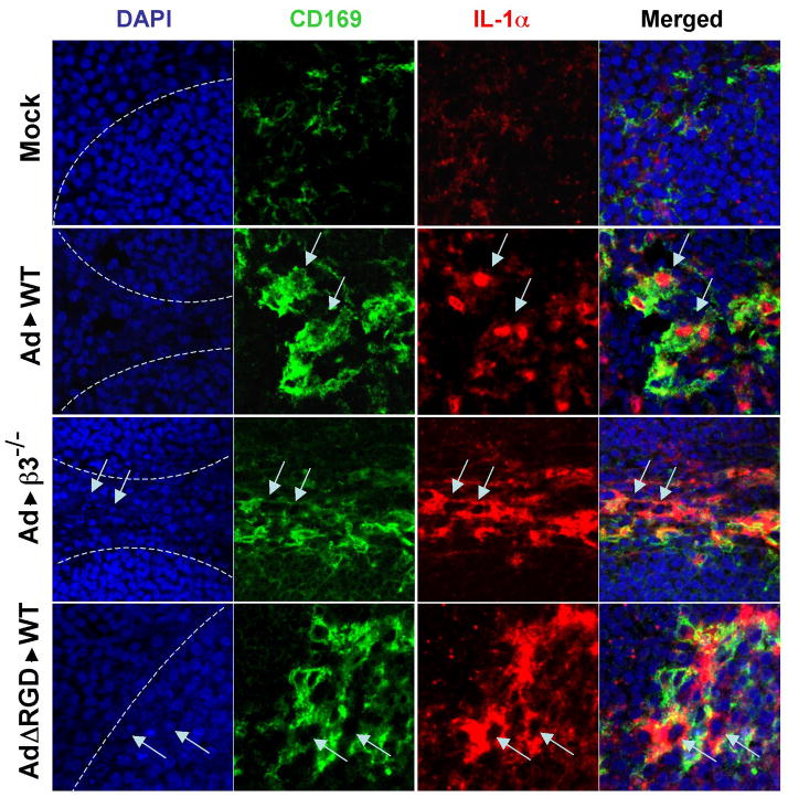
Figure 6. Confocal microscopy analysis of IL-1α translocation into the nuclei of marginal zone macrophages in WT mice and β3 integrin knockout mice injected with Ad, or WT mice injected with AdΔRGD mutant.
Mice were injected intravenously with a high dose of the indicated viruses (1011 virus particles per mouse), and 3 hours later spleens were harvested and sections were prepared and stained with DAPI (blue) to detect nuclei of splenocytes, as well as Abs specific for CD169 (green) or IL-1α (red). Confocal images were obtained using a Zeiss 510 Meta Confocal microscope. The physical border of splenic germinal centers are indicated by punctuate lines. Marginal zone macrophages expressing IL-1α are indicated by arrows. Representative pictures are shown. N=4.

