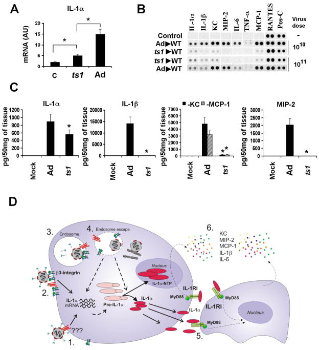
Figure 7. Transcriptional and functional activation of IL-1α in response to ts1 mutant virus and the model of IL-1α-mediated activation of the innate immune response to Ad by macrophages in vivo.
(A) Mice were injected with Ad or ts1 mutant and mRNA levels for IL-1α were analyzed 30 min p.i. N=4. * - P < 0.01. C – mock-infected mice, treated with saline. AU – arbitrary units. (B) Analysis of proteins in spleens of mice 1 hour after Ad or ts1 injection at indicated doses. N=4. Mock – negative control mice injected with saline. Pos-C are dots that show the manufacturer’s internal positive control samples on the membrane. (C) The amounts of pro-inflammatory cytokines and chemokines in the spleens of mice 1 hour after Ad or ts1 injection. N=3. Mock – negative control mice injected with saline.* - P < 0.01. (D) Model of Ad induction of IL-1α that triggers the activation of macrophage innate immune and inflammatory responses in vivo. 1. Ad interaction with a macrophage receptor induces transcription and synthesis of pre-IL-1α. 2–4. β3 integrin interaction with virus RGD motifs triggers intracellular signaling that promotes virus internalization into the cell and endosome rupture, thus, leading to the amplification of IL-1α gene transcription, pre-IL-1α processing, translocation of IL-1α -NTP to the nucleus, and enabling mature IL-1α to initiate IL-1RI signaling. 5. IL-1α -mediated IL-1RI signaling leads to the activation and production of pro-inflammatory cytokines and chemokines, including KC, MIP-2, MCP-1, and IL-6.

