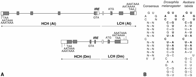
Figure 2. Genomic organization of Ferritin HCH and LCH.
A. Genomic organization of ferritin in A. tabida (At) and in D. melanogaster (Dm) [48]. Introns are represented by lines, and exons by boxes to scale. Within exons, start and stop codons are indicated, delimiting the protein coding regions (shaded gray) and the untranslated regions (white). IRE is shown as dashed box. B. Sequence and secondary structure of the Iron Response Element (IRE) in A. tabida compared to the IRE consensus sequence [32] and the IRE of D. melanogaster [26].

