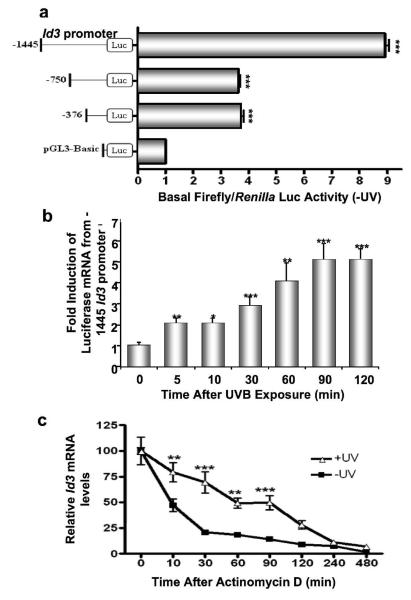
Figure 3.
UVB induces Id3 promoter expression and RNA stability. (a) Different Id3 promoter regions (1445 bp, 750 bp, and 376 bp) were cloned upstream to the firefly luciferase gene, and transiently cotransfected with the pRLTK Renilla vector into immortalized keratinocytes. 24 hours after transfection, cells were either harvested (a), or exposed to UVB and harvested at the indicated time points (b). (a) Luciferase assays were performed and normalized against Renilla luciferase activity to control for transfection efficiency. Fold increase in basal firefly/Renilla luciferase activity is shown for each mutant as a multiple of that obtained using the promoterless pGL3-Basic construct. (b) Immortalized keratinocytes were cotransfected with the Renilla luciferase reporter vector and the −1445 Id3 promoter-luciferase construct. After UVB exposure, RNA was extracted, and luciferase and cyclophilin mRNA levels were measured by QPCR. Fold expression of luciferase/cyclophilin mRNA compared to unirradiated (Time=0) controls are shown as mean ± S.D. of three replicates of a representative experiment. (c) Immortalized keratinocytes were exposed to 480 J/m2 UVB. Actinomycin-D (4 μM) was added 60 minutes after UVB irradiation. Cells were harvested at indicated time points after Actinomycin-D, RNA isolated, and endogenous levels of Id3 mRNA quantified by QPCR. Relative Id3/cyclophilin mRNA levels from UVB-irradiated and unirradiated (Time=0) controls are calculated as mean ± S.D. of three replicates of a representative experiment; essentially the same results were obtained in three independent experiments. One, two or three asterisks represent significance levels of p<.05, .01 or .001, respectively, for each promoter compared to pGL3-Basic (a), or when UVB-exposed is compared to unirradiated controls for each time point (b, c).

