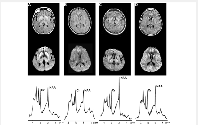
Figure 3.
FLAIR-T2 (top) and DWI (middle), and thalamic 1H-MRS (bottom) from Case #4 with definite sporadic CJD, VV2 (A), Case #2 with definite sporadic fatal insomnia, MM2 (B), Case #18 with possible autoimmune encephalitis (C) and Case #6 with definite sporadic CJD, MM1 (D). On MRI, Case #4 shows increased SI in the striatum and thalamus, Case #2 no SI changes, Case #18 increased SI in the striatum and thalamus and Case #6 increased SI in the striatum and cerebral cortex. On 1H-MRS of the thalamus, all the three prion patients (A, B and D) showed, relative to creatine-phosphocreatine (Cr) a severe reduction of the neuronal marker NAA. The patient without prion disease (C) showed normal thalamic spectrum. ppm = parts per million.

