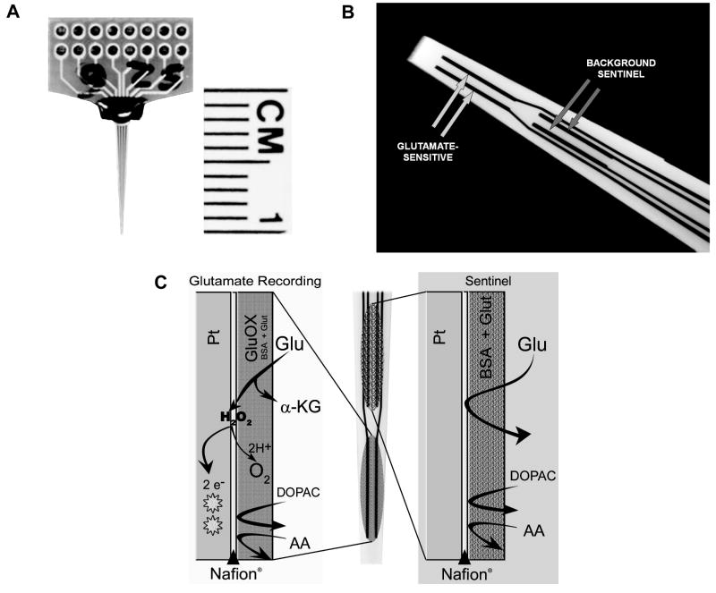
Figure 1.
The glutamate-sensitive microelectrode array (MEA) and the enzyme scheme used in the detection of glutamate. (A) photograph of the MEA with ceramic wafer and recording channels. (B) high magnification of the tip of the MEA illustrating the 2 pairs of recording sites, one pair sensitive to glutamate and the remaining pair as a sentinel for background (non-glutamate derived) signals. (C, left) coatings on the glutamate-sensitive recording sites allowing for the measurement of glutamate-derived current at the Pt microelectrode. (C, right) coatings on the sentinel recording sites for the measurement of background current (see Method for details).

