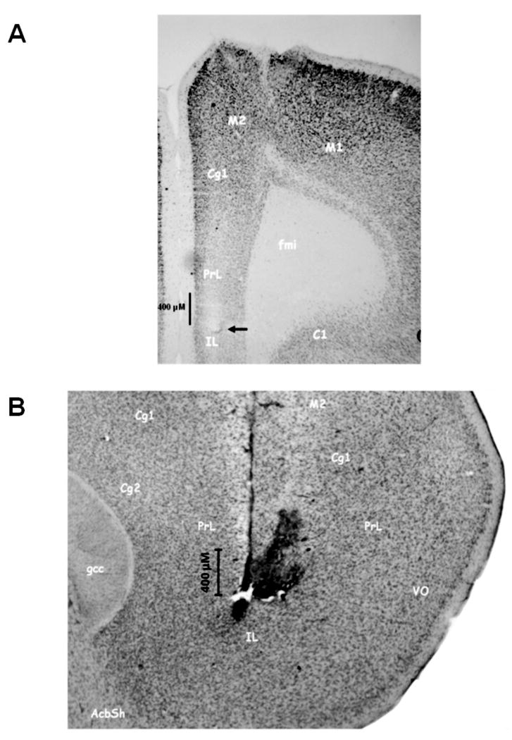
Figure 3.
Photomicrographs illustrating a representative placement of MEA and infusion cannula within prefrontal cortex (PFC). (A) frontal section depicting the position of a microelectrode in the ventral prelimbic region of PFC. The termination of the Pt tip of the MEA is depicted by the arrow above the infra-limbic (IL) cortex. Note the modest tissue disruption produced by the implanted MEA. This is consistent with previous reports from our group (see Rutherford et al., 2007). (B) sagittal section highlighting the relative position of an MEA and the proximity of its infusion cannula. The vertical descent of the MEA can be clearly seen again within pre-limbic (PrL) and infra-limbic (IL) cortex. The modest tissue disruption to the right of the MEA reflects that produced by the infusion cannula and the subsequent infusions of drugs. The close proximity of the end of the infusion cannula and the recording channels of the MEA is evident.

