
Figure 3.
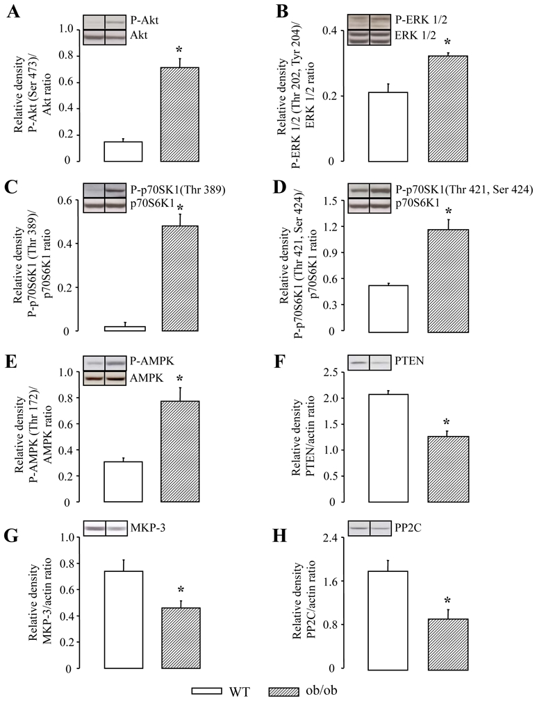
Western blot analysis of Akt and its phosphorylated form at Ser 473 (panel A), ERK 1/2 and its phosphorylated forms at Thr 202 and Tyr 204 (panel B), p70S6K1 and its phosphorylated form at Thr 389 due to the effect of Akt (panel C), p70S6K1 and its phosphorylated form at Thr 421 and Ser 424 due to the effects of ERK1/2 (panel D), AMPK and its phosphorylated form (panel E), PTEN (panel F), MKP-3 (panel G) and PP2C (panel H) in wild-type (WT) and ob/ob mice performed at baseline (i.e., in the absence of ischemia-reperfusion). Values are expressed as mean ± SEM (n=5 per group for WT mice and n=5 per group for ob/ob mice). *p<0.05 vs WT.